


IBS researchers develop high-resolution “ultra-thin” endoscope
Researchers at the Institute for Basic Science (IBS) say they developed a high-resolution holographic endoscope system.
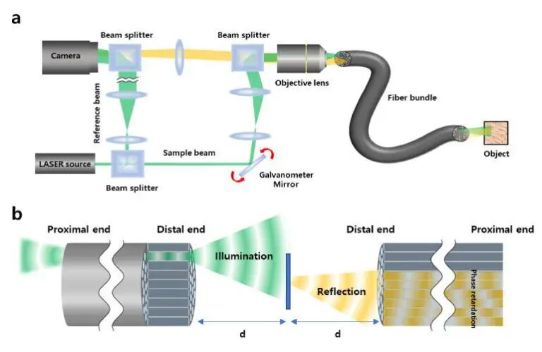
Choi Wonshik, associate director for the Center for Molecular Spectroscopy and Dynamics (CMSD) within IBS, led the team. According to a news release, the researchers overcame the previous limitation of fiber optic endoscopy. They reconstructed high-resolution images without attaching a lens or any equipment to the distal end of the fiber bundle.
The researchers say general endoscopy attaches a camera sensor to the end of a probe using an optical fiber. With camera sensors, the probe thickness increases, making the endoscopy rather invasive. Manufacturing in a thinner form factor can minimize invasiveness and thus patient discomfort. However, a conventional fiber bundle endoscope struggles to perform high-resolution images because the size of the individual fiber cores limits the resolution of the obtained images.
The IBS researchers overcame these limitations by measuring holographic images of the light waves reflected from the object and captured by the fiber bundle. They found they could reconstruct the object image with a microscopic resolution by correcting the phase retardation that occurs by each fiber core.
IBS said the diameter of the endoscope probe totals 350 μm. That offers a thinner option compared to the needle used for hypodermic injection. The researchers obtained images with a spatial resolution of 850 nm — far smaller than the core size of the optical fiber bundle.
They tested the system to image the villi structure of mice. The system proved capable of acquiring high-contrast images, even in biological samples with low reflectivity.
IBS said the researchers believe their application of the new endoscope could improve the way we image the internal structures of the body. This can occur in a minimally invasive manner with little to no discomfort to patients.
Other applications could include microvessels and small airways in the lungs. The researchers said this previously was not feasible. Additionally, it could move beyond the medical field and into inspections of semiconductors and microprocessors.
Article Source: Medical Design Outsourcing