


3D Printing in Dental Prosthodontics, Orthodontics, and Surgery
3D printing technology is making deep inroads in the heavily regulated medical device space. This second blog of a series showcases 3D printed medical devices for prosthodontics, orthodontics, and surgery.
Dentists and dental surgeons have a wide range of methods and treatment solutions for their patients. 3D printing covers many requirements used in more traditional planning methods and medical devices. Scanning the patient is becoming part of all treatment workflows that incorporate 3D printing. After the patient scan, however, workflows differ greatly based on the treatment.
Prosthodontics
Patients who have an indication for prosthodontics (i.e., a prosthetic, a separate piece of non-biological material, for teeth) include those with fractured, greatly decayed, or completely broken/cleaved teeth. These are beyond the scope of fillings due to the larger portion of material requiring replacement by a separately produced and shaped prosthetic. Such replacements include crowns, bridges, and onlays/inlays (partial crowns). In cases where prosthetics are indicated, the clinician and patient decide which material and procedure to proceed with by jointly considering cost, aesthetics, and treatment efficacy/longevity. Once these decisions are in place, the clinician then works in a treatment plan. The first step is a cut to the tooth or teeth in question, and subsequently scan the tooth preparation. An example of a cut preparation is shown in Figure 1.
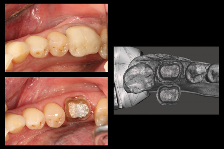
Figure 1: Patient uncut (top left), cut preparation (bottom left), and scanned STL viewed on monitor (right). Image courtesy of Michael Scherer, DMD.
After cutting, scanning captures the 3D structure of the prepared tooth. Using imaging capture software, this is stitched into a 3D surface of the archway or area of interest. In crown planning this can, and in many cases should, include soft tissue like the gingiva. This allows the clinician or technician to plan marginal lines for the produced crown. The scan can then be used to generate a die, which allows for fit to be checked on a produced crown.
Here 3D printing can replace traditional workflow. Rather than creating a die via plaster, a 3D-printed high fidelity version is rapidly produced. It can be printed as a separate piece and fit into a print of the rest of the teeth and jaw, enabling fitment checks between interstitial teeth and the opposing teeth bite pattern. A full workflow of the initial to final patient visit is shown in Figure 2.
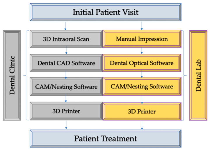
Figure 2: Dental CAD/CAM workflow using 3D printing. Image courtesy of Pillai et al., Polymers.
For crown fabrication, a castable wax resin is printed and used for investment casting via the lost wax technique, as shown in this video. Potential alignment issues associated with a traditional pour-and-shape method are avoided by having the negative (crown form) wax portion printed to spec, and with 20% wax content providing a balance between stability during processing and burn out in oven before ceramic is poured. Some material can even be used for other dental prostheses.
A crown can, with new resins, even be printed directly. Printing to the shape of a crown itself, and finishing with traditional tools in house, allows for quick turnaround and lower cost than traditionally produced crowns. However, the materials are less robust and prone to wear compared to traditional ceramic crowns, requiring replacement sooner.
Planning for Dental Surgery
With the infrastructure in place, alternate resins can be employed to create anatomical models used for implant surgical planning. This includes both rigid and soft tissue material analogues, such as the gingiva and alveolar bone. Being able to view and manipulate both jaws allows the dental surgeon to plan for access to drill the pilot hole for the implant screw base, without needing the patient in-house for these steps. It also provides a tactile alternative to viewing the scans on a computer.
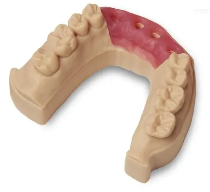
Figure 3: Rigid teeth and jaw analogues, soft gingiva analogue. Image courtesy of Formlabs.
Orthodontics
In addition to using 3D printed jaws for crown checks, they are also used for orthodontics treatment. A crucial step in the workflow of providing traditional metal braces to a patient is placing the base of the brace itself onto each treated tooth. This is traditionally done by placing it close to the intended position, using no position cues beyond the anatomy itself. 3D-printed guides can be used to position and place the braces onto the teeth with measurable accuracy for a full arch of teeth, increasing patient throughput by reducing chair time.
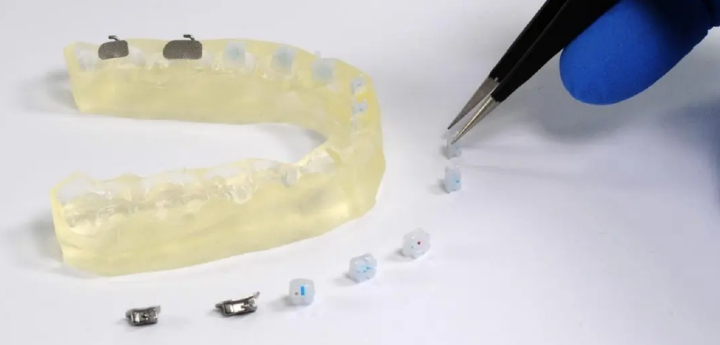
Figure 4: 3D-printed braces tray. Image courtesy of Formlabs.
Conclusion
3D printing is making inroads in dentistry and disrupting traditional approaches through several novel methods and medical devices. It offers the industry more choices for both patients and clinicians for treatment options, which reduce costs while increasing the quality of care provided to patients.
Article source: MPO